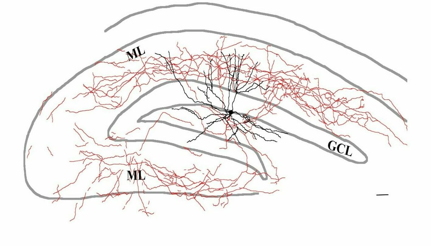
Pyramidal cells, the principal excitatory cells of the hippocampus, are under the control of a remarkably diverse set of inhibitory interneurons. These interneurons show substantial differences in their morphology, physiological behavior (spiking activity) and neurochemical signatures. A specific combination of these three characteristic features allows an interneuron to fill a specific niche within the hippocampal network. Therefore, to better understand the function of a given interneuron type, we need to gain information about all these features.
It is possible to achieve all of these goals using a juxtacellular recording & labelling method that we perform on awake, non-anesthetized, head-fixed mice that are running or resting on a spherical treadmill. To record neuronal spiking activity, we first position a small glass electrode adjacent to the cell membrane, thus recording action potentials without perturbing the cell (i.e. juxtaposing the pipette to the cell membrane). Following recording, an intracellular tracer such as neurobiotin can be delivered specifically to that cell, enabling later morphological reconstruction of the neuron. Subsequently, the labelled cell is further stained for neurochemical markers with immunohistochemistry.
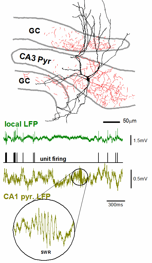
The example on the right shows an interneuron recorded and labeled using the juxtacellular technique. Axons (red) are located in the CA3 region of the hippocampus and in the adjacent blades of the dentate gyrus as well. Morphological and immunohistochemical analysis identified this cell as a unique type of axo-axonic cell that simultaneously controls the axon initial segments of CA3c pyramidal cells and dentate granule cells. Spiking activity (black trace) and simultaneous local field potential (green trace) at high temporal resolution reveals that this cell stops firing long before sharp wave-ripples. For further information, see Szabo GG et al., 2017, Cell Reports.


