By: solteszlab

Jesslyn Homidan is a recent graduate of UC Berkeley, where she received her B.A. in Molecular and Cellular Neurobiology. At Berkeley, she worked as an undergraduate apprentice in Dr. Na Ji’s Lab conducting biophysics research on the thalamus and visual cortex. Jesslyn is excited about her new role as a Research Assistant in our lab.

Aaron Milstein has accepted his new position at Rutgers University. He is an Assistant Professor in the Dept. of Neuroscience and Cell Biology and the Dept. of Neurosurgery at Robert Wood Johnson Medical School, and a resident faculty member at the Center for Advanced Biotechnology and Medicine. As an Instructor mentored by Ivan Soltesz, Aaron developed large biologically-detailed neuronal network models to dissect the circuit components of memory and dysfunction in epilepsy.

The Soltesz Team is wishing him all the best in his new career path!

Barna Dudok received a K99/R00 Career Development Award, entitled “Perisomatic inhibition in epilepsy”. He will study the role of a specific type of GABAergic inhibitory interneuron (cholecystokinin-expressing basket cells) in controlling hippocampal network activity in a mouse model of temporal lobe epilepsy using in vivo calcium imaging.

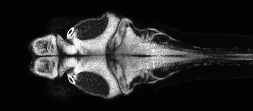
The optical transparency and small brain sizes of larval zebrafish have enabled unprecedented access to single-cell neural dynamics across the whole brain.  With this, we seek to discover novel principles of cellular resolution communication networks that are integral for generating pathological seizure dynamics. Just recently, we have incorporated the chronically epileptic mouse into our analysis. Which is to say, we can now draw insights across evolutionary time, and which will be directly relevant towards translation. Our key advances so far:

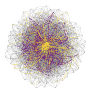
  • Built biologically constrained single-cell whole brain effective connectivity models optimized from experimentally acquired calcium dynamics as a platform for hypothesis testing.
  • With the help of novel clustering algorithms, we discovered a new ‘type’ of neuron – the higher-order hub neuron – which is responsible for destabilizing epileptic networks by facilitating unrestrained excitation downstream.
  • Things are surprisingly similar in chronically epileptic mice! This means we have characterized a previously unrecognized organization of epileptic networks that leads us to predict a new target for seizure control.

Moving forward, we will use our lab’s full-scale data-driven models of hippocampal dentate gyrus and CA1 to dig deeper into the underlying circuit mechanisms responsible for the emergence of higher-order hubs in epileptic networks.
Screen Shot 2020-10-02 at 9.18.30 AM

Lab Members

No members found
April 7, 2020

Octopus Research

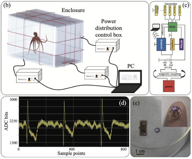
Cephalopods, including the cuttlefish, octopus, and squid, possess one of the most advanced nervous systems among invertebrates. With their advanced nervous systems, cephalopods are able to perform sophisticated behaviors such as navigating in open water to search for food. Yet how their nervous systems accomplish spatial navigation remains completely unknown. On the other hand, much work has been done in mammals, especially the rodent, to reveal the part of their nervous systems underlying spatial navigation. In particular, neurons in the hippocampus exhibit spatially-selective activity that is thought to provide an internal estimate of animals’ current location. This brings up an interesting question: do cephalopods, with their lineage diverges from the vertebrates more than 500 million years ago, evolve a similar or distinct strategy in their nervous systems to solve the navigation problem? To address this question, this project aims to record neuronal activity in the octopus brain while the octopus perform a spatial navigation task. This work has the potential to reveal essential neuronal mechanism underlying spatial navigation, irrespective of different animal species.

Lab Members

Postdoctoral Researcher

Ernie Hwaun

Postdoctoral Researcher

Ernie Hwaun

Ernie completed his PhD in Neuroscience from the University of Texas at Austin. He is interested in how neurons connect with each other to support cognitive functions such as memory. To tackle this problem, Ernie has been using in vivo extracellular recording techniques to obtain neuronal activity while animals perform a memory task. Besides research, Ernie enjoys playing basketball with friends and reading manga.

December 18, 2019

Juxtacellular Recordings

Pyramidal cells, the principal excitatory cells of the hippocampus, are under the control of a remarkably diverse set of inhibitory interneurons.  These interneurons show substantial differences in their morphology, physiological behavior (spiking activity) and neurochemical signatures. A specific combination of these three characteristic features allows an interneuron to fill a specific niche within the hippocampal network. Therefore, to better understand the function of a given interneuron type, we need to gain information about all these features.

It is possible to achieve all of these goals using a juxtacellular recording & labelling method that we perform on awake, non-anesthetized, head-fixed mice that are running or resting on a spherical treadmill. To record neuronal spiking activity, we first position a small glass electrode adjacent to the cell membrane, thus recording action potentials without perturbing the cell (i.e. juxtaposing the pipette to the cell membrane). Following recording, an intracellular tracer such as neurobiotin can be delivered specifically to that cell, enabling later morphological reconstruction of the neuron. Subsequently, the labelled cell is further stained for neurochemical markers with immunohistochemistry.

The example on the right shows an interneuron recorded and labeled using the juxtacellular technique. Axons (red) are located in the CA3 region of the hippocampus and in the adjacent blades of the dentate gyrus as well. Morphological and immunohistochemical analysis identified this cell as a unique type of axo-axonic cell that simultaneously controls the axon initial segments of CA3c pyramidal cells and dentate granule cells. Spiking activity (black trace) and simultaneous local field potential (green trace) at high temporal resolution reveals that this cell stops firing long before sharp wave-ripples. For further information, see Szabo GG et al., 2017, Cell Reports.

Lab Members

Research Scientist

Gergely Szabo

Research Scientist

Gergely Szabo

Gergely is a Basic Life Research Scientist whose main focus is studying the structure and function of hippocampal inhibitory circuitry and its involvement in learning and memory, utilizing techniques such as electrophysiology, optogenetics, and imaging. Gergely received his MS in Biology from Eotvos Lorand University in Hungary and his Ph.D. in Neuroscience from Semmelweis University in Hungary, after which he joined the Soltesz Lab as a postdoctoral fellow.

December 18, 2019

Behavioral Experiments

A major impediment to progress towards reproducible, rigorous preclinical research in epilepsy is that current experimental approaches often require prohibitively time and labor-intensive 24/7 video-EEG monitoring and inherently subjective scoring of seizures by human observers. In collaboration with the Datta lab at Harvard, who showed that complex animal behaviors are structured in stereotyped modules (“syllables”) at sub-second timescales arranged according to specific rules (“grammar”), we are developing a technique employing video imaging with a 3D camera and artificial intelligence (AI)-assisted video analysis to assess epileptic behavior in an automated and unbiased manner.

Lab Members

Instructor

Tilo Gschwind

Instructor

Tilo Gschwind

Focusing on hippocampal network reorganization in temporal lobe epilepsy while tackling inherent problems of decade-old technology to advance epilepsy research, his project in the Soltesz lab provides an optimal opportunity to contribute to the interdisciplinary discourse between the fields of neuroscience and AI.

We use high-density silicon probes in the hippocampus and related circuits to record from single neurons and monitor the local field potential dynamics with high spatiotemporal resolution. When combined with optogenetics, we can “tag” neurons to achieve cell-type specific recordings as mice navigate a cue-rich treadmill while head-fixed or during restraint-free behavior. Optogenetics also enables us to study how specific cells within and outside of the hippocampus control important LFP dynamics, including theta and sharp-wave ripples. We use a variety of electrode layouts, including Neuropixel probes, to cover a range of research questions.

Lab Members

Postdoctoral Researcher

Ernie Hwaun

Instructor

Tilo Gschwind

Postdoctoral Researcher

Ernie Hwaun

Ernie completed his PhD in Neuroscience from the University of Texas at Austin. He is interested in how neurons connect with each other to support cognitive functions such as memory. To tackle this problem, Ernie has been using in vivo extracellular recording techniques to obtain neuronal activity while animals perform a memory task. Besides research, Ernie enjoys playing basketball with friends and reading manga.

Instructor

Tilo Gschwind

Focusing on hippocampal network reorganization in temporal lobe epilepsy while tackling inherent problems of decade-old technology to advance epilepsy research, his project in the Soltesz lab provides an optimal opportunity to contribute to the interdisciplinary discourse between the fields of neuroscience and AI.