In: News
January 1, 2024
Congratulations Assistant Professor Quynh Anh!
In 2024, Quynh Ann became a faculty member and started her own lab in the department of Pharmacology and Vanderbilt Brain Institute, Vanderbilt University, Nashville. QA’s lab aims to bridge our understanding of neural function and dysfunction at the level of molecules, cells, circuits, and behavior. A central focus of the lab is to understand mechanisms of hyperexcitability in the brain. Aberrant firing of neurons underlies many neurological disorders such as epilepsy and a greater understanding of how this dysfunction arises can pave the way for the identification of novel targets for therapeutic intervention. We utilize techniques such as slice electrophysiology, in vivo silicon probe recording, 2-photon imaging, closed-loop optogenetics, EEG recording, and computational modeling in mice for our investigations. The lab has an interdisciplinary group of collaborators, including biochemists and bioengineers who develop tools to aid our basic science investigations as well as clinicians who help translate our basic insights from mouse to humans.
Another Soltesz Lab reunion at AES in the books.

September 1, 2023
Congratulations Assistant Professor Jordan!
In 2023, Jordan became a faculty member and started his own lab in Neurology at Harvard Medical School and the Rosamund Stone Zander Translational Neuroscience Center and F.M. Kirby Neurobiology Center at Boston Children’s Hospital. His lab aims to understand how basic mechanisms that support healthy brain functions become hijacked in epilepsy to drive pathophysiology. His lab’s most recent focus is unravelling how local circuit and large-scale network mechanisms, which normally control memory processes, become substrates for hypersynchronous, pathological activity in epilepsy. To translate their findings, his group develops non-invasive ultrasound approaches to re-tune neural circuits with high spatial and cell type-specific precision.
July 10, 2023
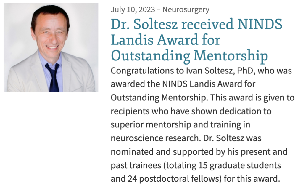
Congratulations, Ivan!
Congratulations to Ivan on being selected for the Landis Award for Outstanding Mentorship.
This annual award is from the National Institute of Neurological Disorders and Stroke (NINDS) at the National Institutes of Health (NIH). The NINDS established this award to emphasize mentorship and encourage faculty to make mentorship a vital component of their career. An incredible achievement and honor!


May 23, 2023
Congratulations, Ryan!

Last week, Stanford Neurosurgery held its first annual Neurosurgery Department Awards. Awards were given to both residents and med students who have shown exemplary work and research in their areas of interest. Ryan Jamiolkowski, who is performing research in our lab, was one of the awardees. Congratulations!
March 1, 2023
Florian – Welcome to Soltesz Lab!
Florian completed his PhD in Neuroscience with Dr. Jean Christophe Poncer at Sorbonne University (Paris). During his PhD, he explored the mechanisms regulating the expression and function of the K+/Cl- transporter type 2 (KCC2) in the brain and investigated the therapeutic potential of targeting this transporter in temporal lobe epilepsy (TLE). He is excited going the lab and will focus more specifically on the mechanisms underlying pathological ensemble activities in the epileptic brain.
February 7, 2023
Balazs – Welcome to Soltesz Lab!
We are thrilled to welcome Balazs in his new role as a Research Assistant in our lab. With two decades of experience in neuroscience, pharmacological research, and development, Balazs is a highly skilled behavioral pharmacologist.
January 1, 2023
Congratulations, Assistant Professor Barna!
In 2023, Dr. Barna Dudok moved to Baylor College of Medicine, Houston, Texas, where he started his new job as McNair Scholar and Assistant Professor at the Department of Neurology. His research is focused on better understanding how GABAergic inhibitory interneurons shape circuit dynamics in healthy brains and in epilepsy. His goal is to identify optimal targets for neuromodulatory intervention and develop cell type-specific strategies for inhibiting epilepsy.
September 1, 2022
Charlotte – Welcome to Soltesz Lab!
Charlotte has been with Stanford since 2021 and joined the Soltesz Lab just recently in September. She is holding a BSc degree in Physiology and Neuroscience from University of California, San Diego. Charlotte is excited about her new role as a Research Assistant in our lab and is currently being mentored by Peter Klein.
Jordan S. Farrell received a K99/R00 Career Development Award, entitled “Dissecting hypothalamic pathways for seizure control”.